The Blastoderm in Chicks During Early Gastrulation
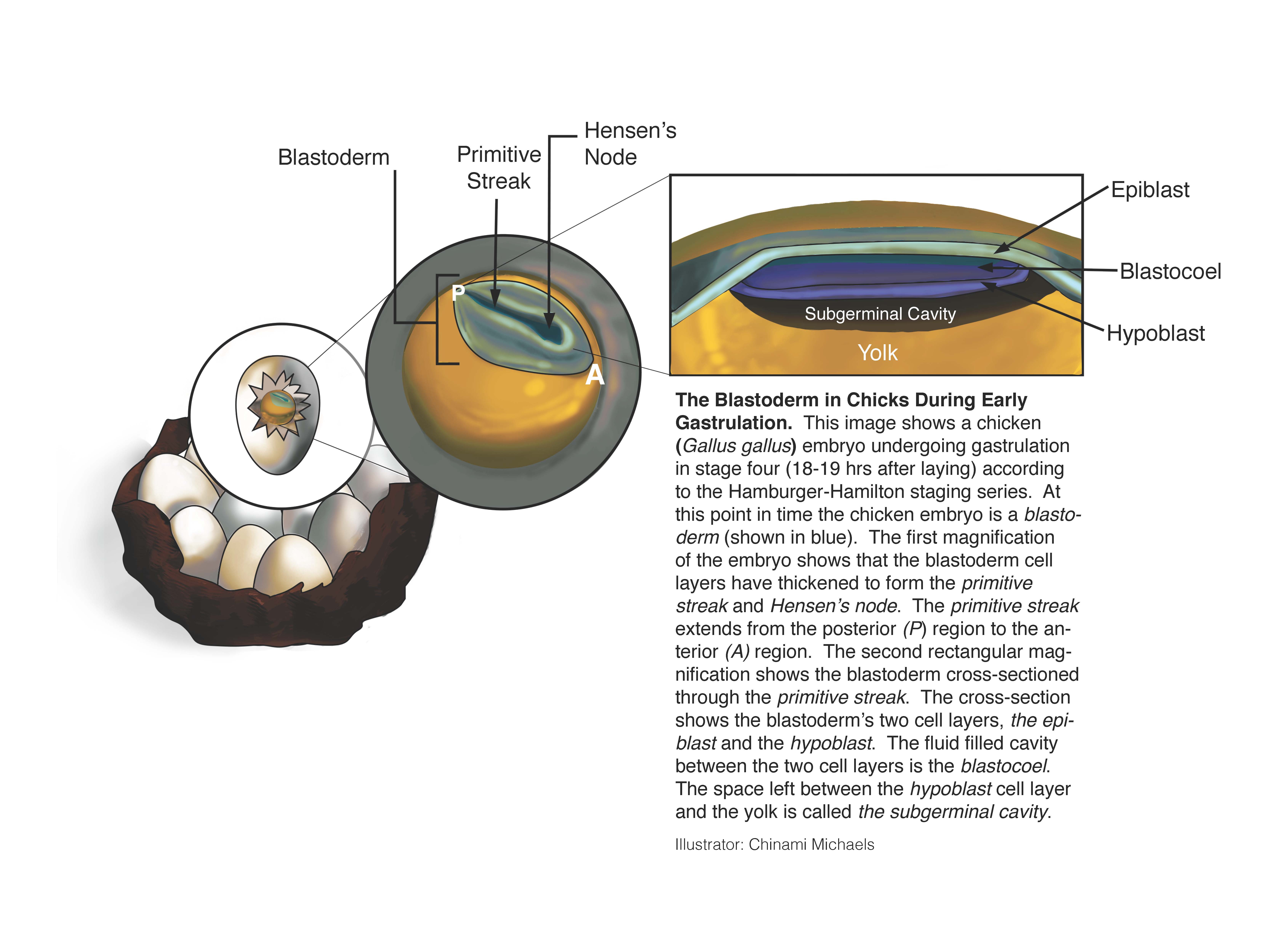
This image shows a chicken (Gallus gallus) embryo undergoing gastrulation in stage four (18-19 hrs after laying) according to the Hamburger-Hamilton staging series. At this point in time the chicken embryo is a blastoderm (shown in blue). The first magnification of the embryo shows that the blastoderm cell layers have thickened to form the primitive streak and Hensen's node. The primitive streak extends from the posterior (P) region to the anterior (A) region. The second rectangular magnification shows the blastoderm cross-sectioned through the primitive streak. The cross-section shows the blastoderm's two cell layers, the epiblast and the hypoblast. The fluid filled cavity between the two cell layers is the blastocoel. The space left between the hypoblast cell layer and the yolk is called the subgerminal cavity.
Keywords
How to cite
Publisher
Handle
Rights
Articles Rights and Graphics
Copyright Arizona Board of Regents Licensed as Creative Commons Attribution-NonCommercial-Share Alike 3.0 Unported (CC BY-NC-SA 3.0)